-
Careers • News • Contact us •
- Login
- Français
 |
Dominic Filion Director |
Team : Mathew Duguay, Research Assistant
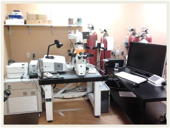
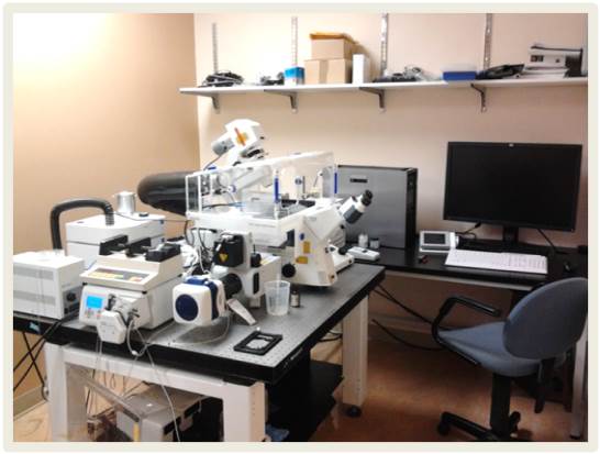
The Microscopy and Imaging platform is equipped with optical microscopes to acquire images or videos of various live or fixed samples. Several detection systems allow these acquisitions to be made. In addition, different software programs are used to process the acquired images and thus obtain digital data such as cell quantities, relative fluorescence intensities, surface measurements, displacement speeds, etc. Our staff aims to obtain uniform images and videos whose quality allows their analysis by automatic computer programs. Finally, tour team develops various customized analysis tools.
|
|
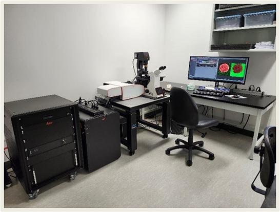
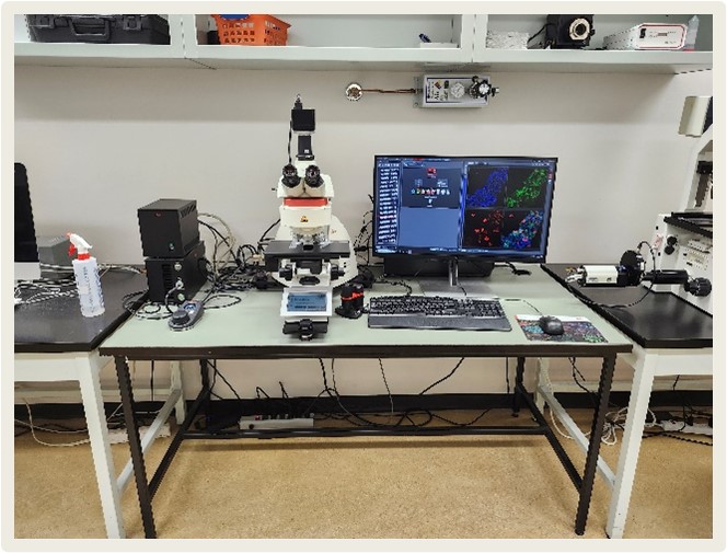
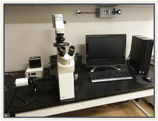
Confocal Stellaris-stedThe Leica Stellaris-STED confocal microscope is equipped with laser sources for excitation at 405 nm, as well as a tunable white laser ranging from 485 to 685 nm. The system also enables super-resolution STED microscopy using a depletion laser at 775 nm. This technique requires specific fluorescent markers, but there are options available that allow double labeling and, in some cases, even triple labeling. Under optimized conditions, it is possible to resolve details below 50 nm. The system includes four adjustable detectors covering the range from 410 to 750 nm, with the capability to measure fluorescence lifetimes (TauSeparation). |
|
|
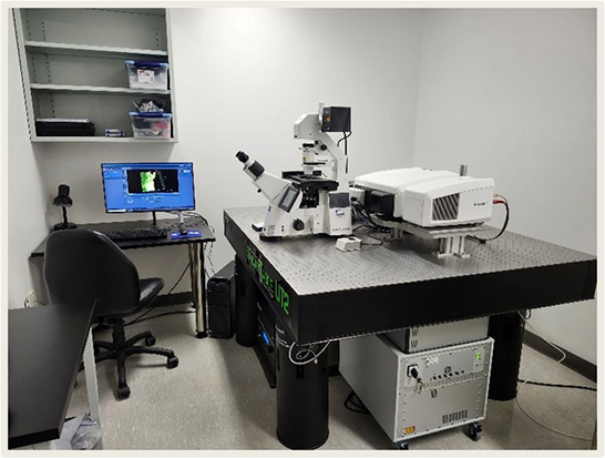
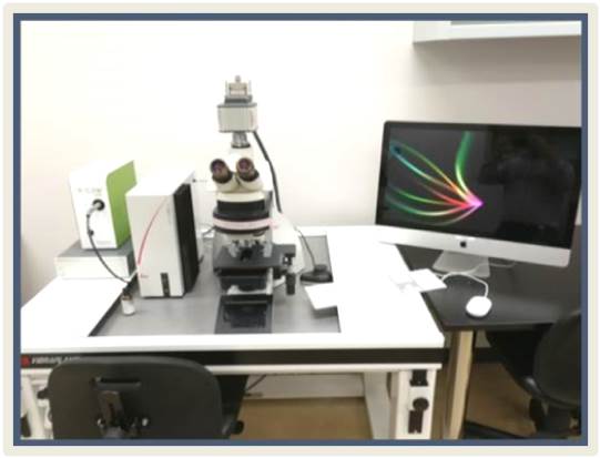
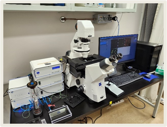
Confocal LSM980 – Mutliphoton – AiryScan2The Zeiss LSM980 multiphoton confocal microscope with AiryScan2 is equipped with standard laser sources at 405, 488, 568, and 639 nm, as well as a tunable pulsed laser ranging from 680 to 1080 nm for multiphoton microscopy. For detection, the system includes three adjustable PMT detectors covering the range from 400 to 850 nm. It also features an AiryScan2 detector, which enables a resolution of approximately 90 nm, using the same markers as standard confocal microscopy. Thanks to this detector, it is also possible to achieve an acquisition speed of 25 fps while maintaining confocal resolution at 512 × 512 pixels. |
|
|
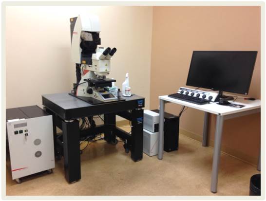
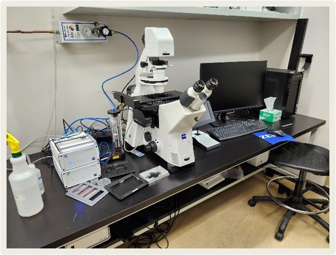
Confocal SP8The LEICA SP8 confocal microscope is a straight microscope equipped with 3 PMTs (Photo-Multiplyer Tubes or photo multiplier tubes) of which two are used for fluorescence and the other detector for transmitted light and one (HyD) hybrid detector (using another principle to detect photons whose sensitivity is increased compared to standard PMTs). The fluorescence emission signals are refracted by a prism and then directed to the detectors. This principle allows a very precise adjustment to separate the signals according to their emission wavelength. This microscope is also equipped with a motorized XYZ stage and includes 4 laser lines for excitation (405, 488, 555 and 638 nm). |
|
|
Confocal LSM 700The confocal microscope LSM700 of the company ZEISS is an inverted microscope equipped with 3 PMT (Photo-Multiplyer Tubes or photo multiplier tubes) of which two are used for fluorescence and the other detector for transmitted light. The signals are separated by a variable dichroic so that the part of the signal whose wavelengths are longer than a given value goes to one PMT while those that are shorter go to the other PMT. This microscope is also equipped with a motorized XYZ stage and includes 4 laser lines for excitation (405, 488, 555 and 638 nm). |
|
|
Confocal LSM 710The confocal microscope LSM710 of the company ZEISS is an inverted microscope equipped with 4 PMTs (Photo-Multiplyer Tubes or photo multiplier tubes) of which three are used for fluorescence and the other detector for transmitted light. The signals are separated by a diffraction grating before being spatially separated by prisms and caches before being directed towards the PMTs. This microscope is also equipped with a motorized XYZ stage and includes 4 laser lines for excitation (405, 488, 555 and 638 nm). |
|
|
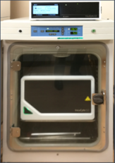
Spinning disk Confocal (rotary disc confocal)The confocal rotary disc microscope from ZEISS is an inverted microscope equipped with a Yokogawa CSU-1 module. This microscope is also equipped with a motorized XYZ stage and an incubator to maintain a stable physiological environment at controlled temperature, percentage of CO2 and O2. There are 4 laser lines for excitation (405, 488, 561 and 639 nm). The system also allows the excitation to be manipulated to perform transitions or photonic damage. |
|
|
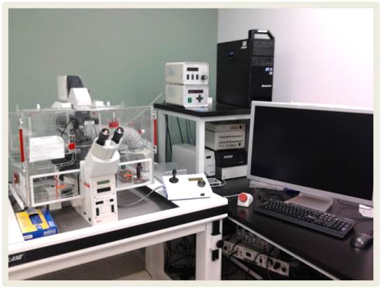
LightSheet Z.1 MicroscopeThe LightSheet Z.1 microscope enables 3D acquisition of specimens larger than 1 cm³ with an XY resolution of approximately 1 µm. The excitation laser sources include 405, 445, 488, 515, 561, and 638 nm.The system is also equipped with two cameras, allowing simultaneous acquisition of two signals for faster imaging. |
|
|
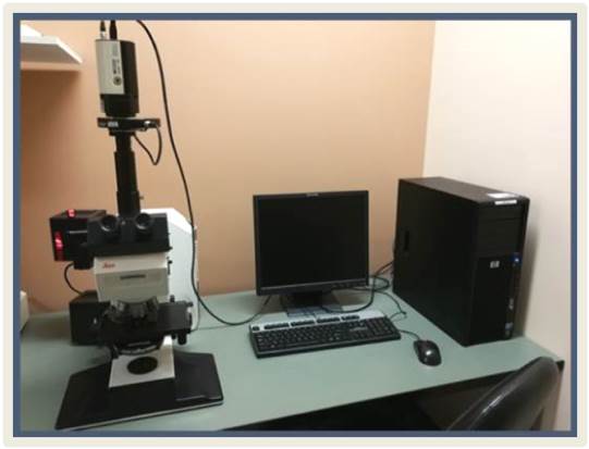
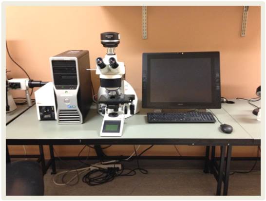
DM6The DM6 microscope of the LEICA company is a straight microscope equipped with a motorized XYZ stage allowing the acquisition of mosaic images. A full LAS X software license allows you to use a multitude of functions. Two cameras are installed on this microscope. The first camera, the ORCAflash 4.0 V.2 from Hamamatsu, a high resolution monochromatic camera (2048 x 2048 pixels of 6.5 x 6.5 μm) allows the acquisition of images of fluorescent probes. This system is equipped with 6 filter cubes (Dapi, CFP, YFP, GFP, Cy3 and Cy5) whose light source is of LED type, model X-Cite of the company Lumen Dynamics. The second camera, DFC480 from Leica, allows the acquisition of colour images. |
|
|
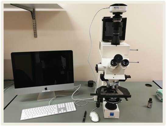
DM6BThe Leica DM6 B microscope is an upright microscope equipped with an XYZ motorized stage, enabling mosaic image acquisition. Image acquisition is performed using the LAS X software. This system includes five filter cubes (DAPI, GFP, Cy3, Texas Red, and Cy5) with an LED light source. It is also equipped with a Leica K5 sCMOS camera, offering a resolution of 2048 × 2048 pixels and a quantum efficiency greater than 80%. |
|
|
DM6000The DM6000 microscope from LEICA is a straight microscope equipped with an ORCA-ER C-4742 camera from Hamamatsu, a monochromatic camera, with a resolution of 1344 x 1048 pixels allows the acquisition of images from fluorescent probes. This system is equipped with 6 filter cubes (Dapi, CFP, YFP, GFP, Cy3 and Cy5) whose light source is of LED type, model X-Cite 120LED Boost of the Excelitas company. |
|
|
DM5500BThe DM5500B microscope from the LEICA company is a straight microscope equipped with a Retiga EXi camera, a monochromatic camera, with a resolution of 1344 x 1048 pixels allows the acquisition of images from fluorescent probes. This system is equipped with 4 filter cubes (Dapi, GFP, Cy3 and Cy5) whose light source is HBO type with mercury short arc. |
|
|
DM4000The DM4000 microscope from the LEICA company is a straight microscope equipped with a Retiga EXi camera, a monochromatic camera, with a resolution of 1344 x 1048 pixels allows the acquisition of images from fluorescent probes. This system is equipped with 6 filter cubes (Dapi, CFP, YFP, GFP, Cy3 and Cy5) whose light source is of LED type, model X-Cite 120LED Boost of the Excelitas company. |
|
|
DMRBThe DMRB microscope from the LEICA company is a straight microscope equipped with a Retiga EXi camera, a monochromatic camera, with a resolution of 1344 x 1048 pixels allows the acquisition of images from fluorescent probes. This system is equipped with 4 filter cubes (Dapi, GFP, Cy3 and Cy5) whose light source is HBO type with mercury short arc. |


|
|
Timelapse CAYThe interval acquisition microscope known as Timelapse CAY allows the acquisition of videos of live samples that can be viewed in accelerated mode. This microscope, surrounded by an incubator whose environment controls temperature and CO2 concentration, is composed of a LEICA DMIRE2 stand, a motorized MS-2000 controller board from ASI Imaging (Applied Scientific Instrumentation). The software managing and communicating the various components is Volocity 6.0 from Perkin Elmer. |
|
|
Timelapse CHAThe interval acquisition microscope known as Timelapse CHA allows the acquisition of videos of live samples that can be viewed in accelerated mode. This microscope, surrounded by an incubator whose environment controls temperature and CO2 concentration, is composed of a LEICA DMIRE2 stand, a motorized MS-2000 controller board from ASI Imaging (Applied Scientific Instrumentation). The software managing and communicating the various components is Volocity 6.0 from Perkin Elmer. |
|
|
Observer 7 Timelapse MicroscopesThese two twin Observer 7 microscopes from Zeiss are equipped with incubation systems that allow temperature control. One of the systems also provides CO₂ regulation. Both microscopes feature DefiniteFocus3 technology, which maintains sample focus throughout time-lapse acquisitions. Each system is equipped with a motorized stage, enabling multi-point imaging. Excitation sources are provided by the Colibri 5 module, which includes four LEDs centered at 375, 475, 555, and 630 nm. The systems are also equipped with Axiocam 820 mono cameras, offering a resolution of 4512 × 4512 pixels. |
|
|
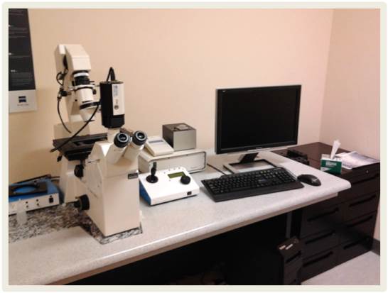
Axiovert S100TV Inverted MicroscopeThe Axiovert S100TV inverted microscope from ZEISS is equipped with a motorized MS-2000 controller board from ASI Imaging (Applied Scientific Instrumentation). Different racks are available to deposit kneaders of various sizes or multi-well plates. The Retiga EXi camera is used to acquire monochromatic images of samples. Several manual filter cubes are available. |
|
|
DMIRE Inverted MicroscopeThe LEICA DMIRE microscope allows the observation of different samples in transmitted light or by fluorescence. Several manual filter cubes are available. If required, images can be acquired by moving a Retiga EXi camera from a nearby system. |
|
|
IncuCyte MicroscopeThe IncuCyte microscope is a high-throughput screening timelapse imaging system, from the company Sartorius. It is mostly used to acquire images of multi-well plates. This microscope can be adapted to most types of well plates. The system acquires phase contrast images and 2 types of fluorescence (green and red). These images are accumulated in a database and can then be analyzed in a completely automated way to perform analyzes of cell counts, intensity, etc. The IncuCyte software can also acquire "scratch essay" type experiments. It is ideal for looking at cell growth curves and variation of fluorescence intensity over time. |
|
|
Straight Microscope DM4000B – OsteomeasureThis DM4000B straight microscope from Leica is equipped with a DP72 camera from Olympus. Osteomeasure software from OsteoMetrics is installed there. This software allows to obtain measurements such as surfaces, perimeters, number of different elements which compose the images. These measurements are generally obtained following drawings made by the user using a touch screen. |
|
|
Straight Axiophot MicroscopeThis straight Axiophot microscope from ZEISS allows you to take photos of histological slides using a Micropublisher camera from Q-Imaging. |
|
|
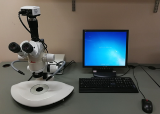
Dissecting microscope MZ12This MZ12 dissecting microscope from LEICA is equipped with a Lumenera Infinity3 camera allowing to take color images with a resolution of 2752 x 2192 whose pixels have a size of 4.54 x 4.54 μm. |
|
|
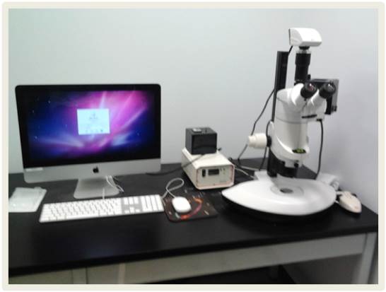
MZ16FA MicroscopeThis MZ16FA dissecting microscope from LEICA is equipped with a Leica DFC350 camera to take colour images on a MAC environment. The software used for image acquisition is a basic Leica capture software. This microscope is also equipped with a filter to visualize the fluorescence of the samples. The light source for fluorescence is of the HBO type with mercury short arc. |
Contact the
Microscopy and Imaging services
110, ave des Pins West,
Montréal (Québec) Canada
H2W 1R7
514 987-5500
Partners

Research
Platforms
Student life
Admissions
The Institure
- A word from the president
- Our impact
- Our Legacy
- Governance
- Management Committee
- IRCM Awards
- Annual reports
Events
The Clinic
© Montreal Clinical Research Institute, Année.All rights reserves. | Privacy policy | Terms of use | Web site by Agence Riposte